Cerebral Ventricle, Ventricular Anatomy, Third Ventricle, Fourth Ventricle, Lateral Ventricle, Cerebral Aqueduct, Cerebral Choroid Plexus, Aqueduct of Sylvius, Middle Foramen of Magendie, Lateral Foramena of Luschka, Cerebrospinal Flow, Subarachnoid Cistern, Glymphatic System
- See Also
- Definitions
- Cerebrospinal Fluid (CSF)
- Central Nervous System floats in a nutrient rich, clear cerebrospinal fluid
- CSF typically contains no white or Red Blood Cells (WBCs or RBCs) and low levels of Protein
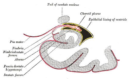
- CSF is produced in the Choroid plexus of fluid filled chambers known as ventricles
-
Choroid Plexus
- Produces cerebrospinal fluid in the walls of the Cerebral Ventricles
- Glymphatic System
- CSF drainage network composed of glial cells and lymphatic-like channels
- Clears waste products (tau Protein, amyloid beta Protein)
- Stimulated by slow rhythmic Norepinephrine release (by locus ceruleus) during non-REM Sleep
- Rhythmic Norepinephrine release is disrupted with impaired glymphatic drainage
- Sleep aids (e.g. Z-Drugs)
- Decreased clearance and longterm Dementia risk
- Traumatic Brain Injury
- Persistent Norepinephrine elevations (may be countered with alpha and Beta Blockers)
- Result in Protein accumulation, cerebral edema, cell death
- Sleep aids (e.g. Z-Drugs)
- References
- Sacchetti (2026) Glymphatic System in Traumatic Brain Surgery, EM:Rap, 10/5/2026
- Dagum (2026) Nat Commun 17(1):715 +PMID: 41593094 [PubMed]
- Zare (2026) Brain Behav 16(4):e71374 +PMID: 41981905 [PubMed]
- Anatomy
- Cerebrospinal Spinal Fluid Pathway
- Images
-
General
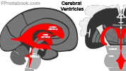
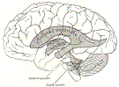
- CSF is produced in the Choroid plexus of the two Lateral Ventricles
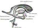
- CSF flows from Lateral Ventricles via foramina to the central Third Ventricle
- From the central Third Ventricle to the Aqueduct of Sylvius and then the Fourth Ventricle
- CSF flows into the subarachnoid space and into the Cerebral Sinuses
- CSF returns to the venous system via reabsorption at the arachnoid villi (esp. in Superior Sagittal Sinus)
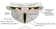
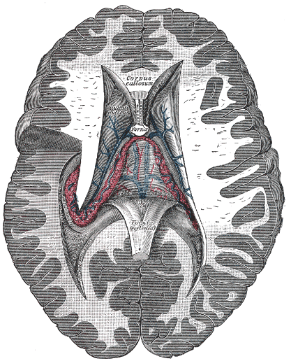
- Lateral Ventricles (left and right)
- Most superior ventricle
- Cerebrospinal fluid originates here (and in the third and Fourth Ventricles)
- Drains via the interventricular foramina (foramina of monro) into the Third Ventricle
- Third Ventricle (midline)
- Drains via the Aqueduct of Sylvius into the Fourth Ventricle
- Fourth Ventricle (midline, posterio fossa)
- Drains out of the intracranial cavity into the subarachnoid space via three openings
- Middle Foramen of Magendie (midline) openings
- Lateral Foramena of Luschka (right and left) openings
- Subarachnoid Space
- CSF flows into the subarachnoid space
- CSF drains into the Superior Sagittal Sinus via arachnoid villi or granules (small holes in the sinus wall)
- Subarachnoid Cistern
- Wider pockets of subarachnoid space and cerebrospinal fluid
- Largest cistern is in the lumbar region (below the L2 Vertebrae)
- Allows for safer Lumbar Puncture

- Pathophysiology
-
Hydrocephalus
- Occurs when CSF flow is obstructed, leading to expansion of the Lateral Ventricles
-
Subarachnoid Hemorrhage
- Ruptured Cerebral Aneurysm results in CSF Red Blood Cells
- With time, yellowing develops of the spinal fluid (Xanthochromia)
- Anatomy
- Ventricles
-
-
 Lewis (1918) Gray's Anatomy 20th ed (in public domain at Yahoo or BartleBy)
-
 Lewis (1918) Gray's Anatomy 20th ed (in public domain at Yahoo or BartleBy)
-
 Lewis (1918) Gray's Anatomy 20th ed (in public domain at Yahoo or BartleBy)
-
 Lewis (1918) Gray's Anatomy 20th ed (in public domain at Yahoo or BartleBy)
-
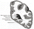 Lewis (1918) Gray's Anatomy 20th ed (in public domain at Yahoo or BartleBy)
-
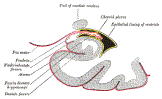 Lewis (1918) Gray's Anatomy 20th ed (in public domain at Yahoo or BartleBy)
-
 Lewis (1918) Gray's Anatomy 20th ed (in public domain at Yahoo or BartleBy)
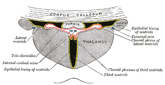


- References
- Gilman (1989) Manter and Gatz Essentials of Neuroanatomy and Neurophysiology, Davis, p. 232-7
- Goldberg (2014) Clinical Neuroanatomy, Medmaster, p. 6-15
- Netter (1997) Atlas Human Anatomy, ICON Learning, p. 102-3