Epidural Hematoma, Epidural Hemorrhage
- See Also
- Epidemiology
- Ages 2 to 60 years
- Dura matter adheres more tightly to skull outside these age ranges, and prevents blood accumulation
- Epidural Hematoma occurs in ~8% of Traumatic Brain Injury (worldwide)
- Often coexists with other CNS Hemorrhage (e.g. Traumatic Subarachnoid Hemorrhage, Subdural Hematoma)
- Pathophysiology
- Epidural Hematoma results from Hemorrhage and blood accumulation between the skull and Dura Mater
- Associated with a Temporal Bone or parietal bone Skull Fracture in 75% of cases
- Involved vessels
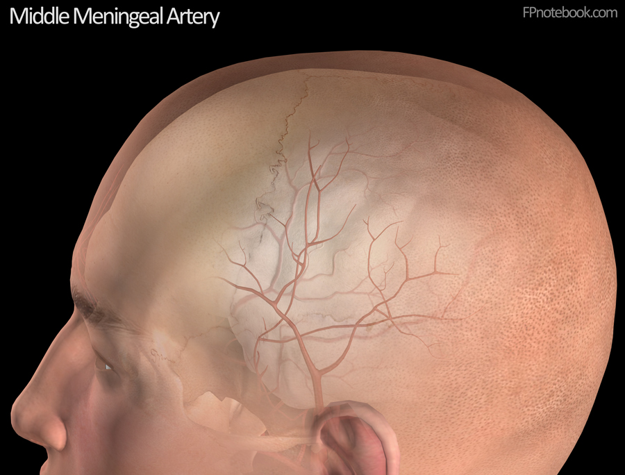
- Middle meningeal artery accounts for 50% of surrgical cases
- Middle Meningeal Artery rupture
- Middle Meningeal Artery rupture
- Other sources
- Dural venous sinuses (40% of surgical cases)
- Middle meningeal artery accounts for 50% of surrgical cases
- Symptoms
- Timing of presentation depends on bleeding source (most present <24 hours)
- Arterial Epidural Hematomas (e.g. middle meningeal artery) develop rapidly (within hours)
- Dural Sinus Epidural Hematomas develop more slowly
- Headache
- Nausea and Vomiting
- Nuchal Rigidity
- Signs
- Pathognomonic Presentation
- Classic presentation occurs in only 20% of patients
- Loss of consciousness
- Period of lucency interspersed between 2 distinct periods of LOC
- Variably present and variable timing
- Absent in most cases, in which patient remains comatose without period of lucidity
- Loss of consciousness
- Signs
- Transtentorial bleed findings
- Contralateral Hemiparesis
- Loss of consciousness eventually occurs
- Ipsilateral fixed and dilated pupil (Cranial Nerve III palsy) in 85% of cases
- Heralds impending Cerebral Herniation
- Imaging
- CT Head
- Focal bleeding that does not typically cross Sutures
- Contrast with subdural which can extend fully anterior to posterior)
- Often in territory of middle meningeal artery (50% of cases)
- Convex "lens" (biconvex) appearance on CT
- Contrast with Subdural Hematoma with concave crescent facing inward (and convexity facing externally)
- Outside the dura, and therefore follows the inner skull surface
- Blood dissects between the skull and the tightly adherent dura
- Findings of continued bleeding
- Swirl sign or active extravasation may be seen when IV contrast is used
- Approximate Hematoma Volume calculation (ABC/2, elipse volume calculation)
- Precaution
- More accurate volume calculations may be done with manual tracing of the Hematoma
- Axial Slice (Transverse Plane): Identify CT slice in which Epidural Hematoma size is maximal
- Coronal Slice (Coronal Plane): Identify CT slice in which Epidural Hematoma size is maximal
- Volume calculation
- Elipse volume = A * B * C /2
- Precaution
- Location of Epidural Hematoma
- Supratentorial
- Superior to the tentorium cerebelli (dural fold marking the upper border of the potserior fossa)
- Infratentorial
- Smaller confined space at higher risk of Brainstem Herniation than supratentorial Hematomas
- Surgical evacuation is performed at lower Hematoma volumes than supratentorial
- Supratentorial
- Other important characteristics of Epidural Hematomas
- Maximum thickness of Epidural Hematoma
- Midline shift
- Mass effect on adjacent structures
- Precautions
- Epidural Hemorrhage may be rapidly fatal
- Mortality is 5 fold higher in delayed diagnosis
- Evaluation
- See Trauma Evaluation
- See Head Injury
- Management
- See ABC Management
- See Management of Severe Head Injury
- See Increased Intracranial Pressure in Closed Head Injury
- Rapid assessment and management is key
- Emergent Neurosurgical Consultation
- Emergent decompression in the Emergency Department
- Indicated in imminent Cerebral Herniation (Ipsilateral fixed and dilated pupil) and delay to neurosurgery
- See Skull Trephination
- Neurosurgical decompression indications
- Epidural Hematoma width >15 mm
- Epidural Hematoma volume >30 ml (cm^3)
- Midline shift >5 mm
- Poor mental status (GCS <8)
- Impending brain Herniation
- Bullock (2006) Neurosurgery 58(3 suppl): S7-15 [PubMed]
- References
- Abuguyan (2024) Crit Dec Emerg Med 38(7): 4-11
- Broder and Lee (2026) Crit Dec Emerg Med 40(6): 24-7
- Dreis (2020) Crit Dec Emerg Med 34(7):3-21